surgery

Sea Turtle Surgery

If you know Dr. Grafinger at all, you probably know that he really enjoys working with exotics; the staff here at TVRH enjoys that he enjoys them.  We see hawks and owls and beavers and otters from the wildlife rehabilitator group CLAWS. We work on tigers and lions and caracals and Geoffrey’s cats from the rescue organization The Conservators’ Center (some of their photos are on the wall in his exam room).  And recently, some of us got to help out with surgery on a Kemp’s Ridley sea turtle and a green moray eel.

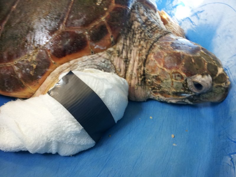
Dr Grafinger has performed surgery on several sea turtles (the previous one from the Karen Beasley Sea Turtle Rescue and Rehabilitation Center, seen here in a previous blog post), so when the North Carolina Aquarium on Roanoke Island  needed help with an injured turtle, they gave him a call.

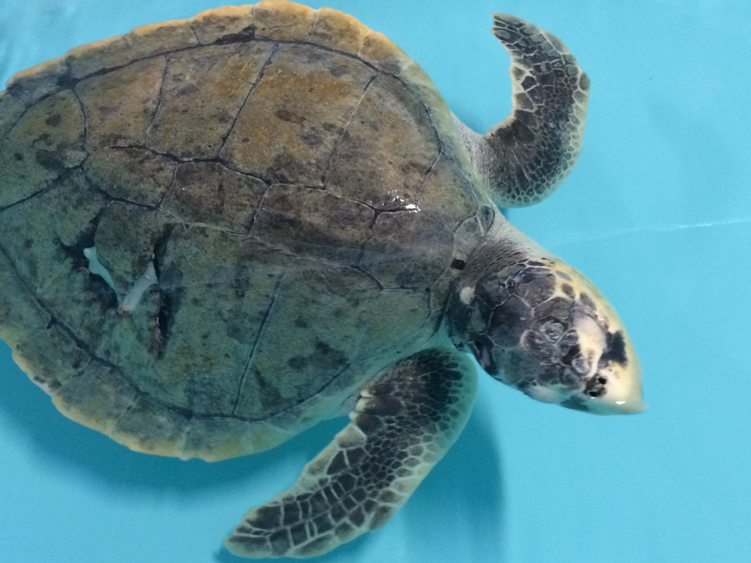
In November of 2014, the Sea Turtle Assistance and Rehabilitation (STAR) Center at the Aquarium obtained a sub-adult Kemp’s Ridley turtle that they nicknamed Finn.  Finn had partially healed wounds on his carapace (shell), a puncture wound to the head, and a swollen left front flipper (all possibly inflicted by a boat propeller).  The doctor and staff at the aquarium observed that Finn was lethargic, usually kept both eyes closed, and he was not eating. 

The wound on the carapace was healing well and required no further treatment.  Finn received extensive care for the wound on his head, and bits of bone and necrotic tissue were removed at each cleaning; he was treated with antibiotics, pain medications, and force feedings.

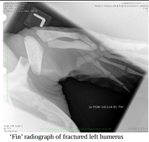
Radiographs and a CT indicated that Finn’s head tilt was probably due to trauma and pain (not brain damage) and that the humerus (the upper “arm” bone of his flipper) was fractured into three pieces, with evidence of bone lysis (the bone was dissolving) as well as possible infection of the bone.  This flipper issue was what Dr Grafinger was there to try to fix.  Oy!

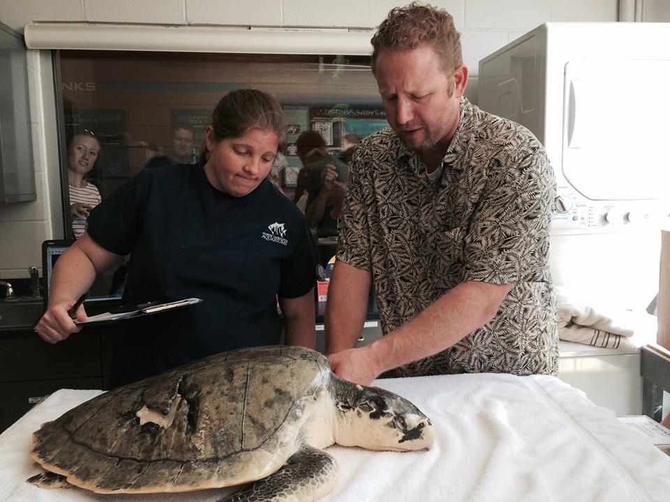
By the time our TVRH team made the road trip to Manteo, Finn had been undergoing minor procedures and receiving wound care for five months.  He was doing some eating on his own and gaining weight, but his neck and flipper wounds were still producing smelly, necrotic material.  Dr Grafinger and the aquarium team agreed on a plan of attack, and then it was time to try to repair this amazing animal.

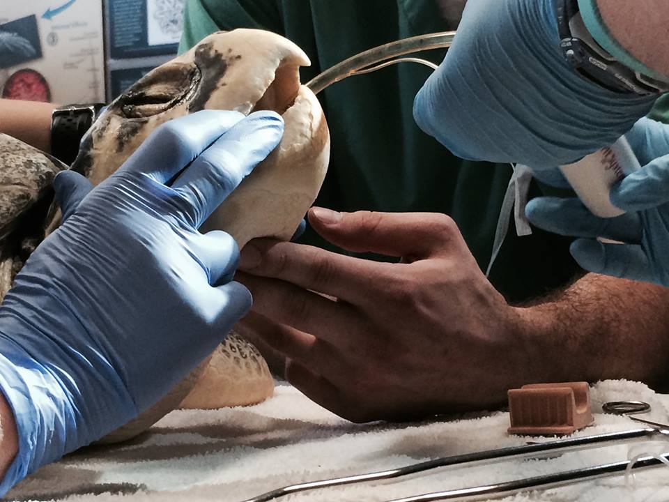
After the experienced aquarium team sedated, intubated, and anesthetized Finn, Dr Grafinger’s surgery assistants (Samantha and Rafe) prepared the site.  Dr Grafinger then made the initial incision that would allow him to access the affected area of the humerus without damaging vital nerves or blood flow.  What he found added an extra degree of complication to Finn’s surgery – the center piece of bone that had broken loose was dead (the source of the odor and infection).

That dead piece of bone was nearly a third of the length of the humerus, so Dr Grafinger had to find a way to stabilize the remaining bone and reduce that open space.  Pins and plates (hardware that’s often used in orthopedic surgeries) just weren’t going to work, so he had to come up with a new plan.

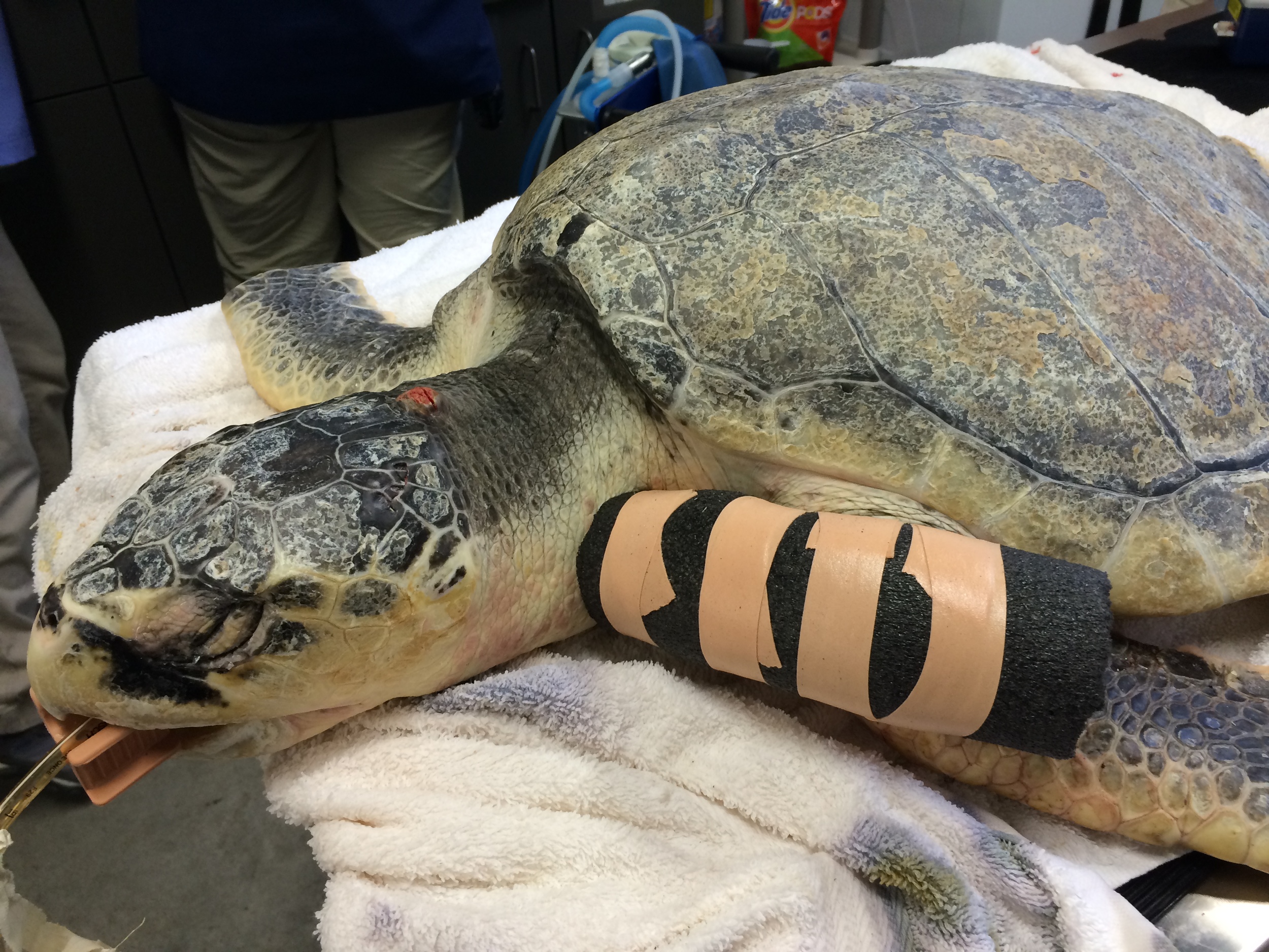
What he used was external fixation.  External fixation is a method of stabilizing bone and soft tissue at a distance from the original injury;  Dr Grafinger would drill into healthy bone, pass metal pins through those drilled holes, and then pass them back out of the body. 

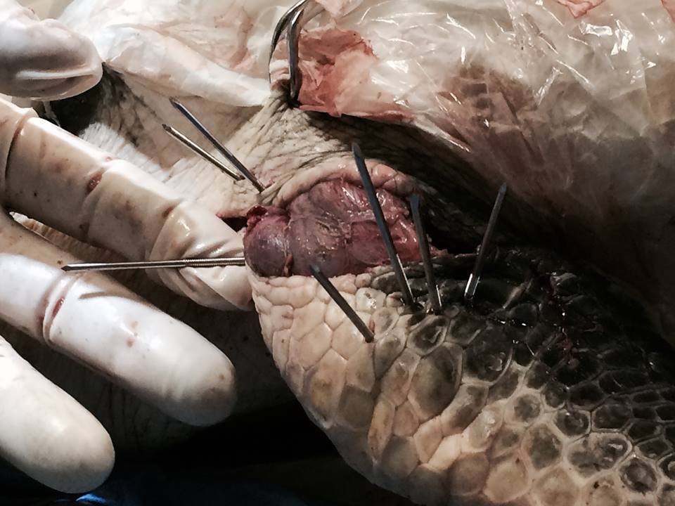
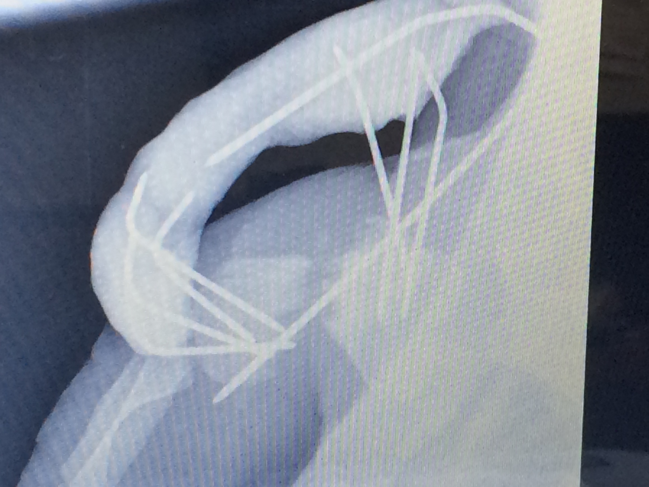
Once the pins were placed to his satisfaction, a fast-hardening epoxy putty was used on the outside of the incision to keep those pins from moving, allowing the bone to heal.  Then the entire putty arrangement was protected by using pipe insulation in case Finn managed to bump it in his tank. 

Dr Grafinger managed to get those distant bone edges closer together, but Finn will still need to do some significant remodeling (new bone formation) to close that open space.  Luckily, the aquarium’s staff veterinarian has found that sea turtles are very good at that sort of repair, so we have high hopes for Finn’s ultimate outcome.

We don’t know yet if Finn will thrive, or even survive; he suffered a lot of damage.  But with the support and knowledge of the aquarium team and Dr Grafinger’s best effort to repair that flipper, we’re all hoping that Finn can go back into the wild at some point in the future.  You can bet that when that day comes, the TVRH team will be there to see it, cheering him on.

IMG_3851.JPG

Meet Oscar

Oscar is looking great 2 ½ weeks after his surgery. I was ecstatic to see him recover so well after surgery.

Oscar is looking great 2 ½ weeks after his surgery. I was ecstatic to see him recover so well after surgery.

Oscar, a 3 year old male Doberman Pinscher, was running in the back yard with the owners when suddenly he jumped on pile of wooden sticks. The owners were surprised and horrified to see a large wooden stick coming out of the right chest wall when he jumped out of the pile. The stick was sticking out of his right chest wall and for a small period of time Oscar was suspended in the air only supported by the stick when the stick broke. Oscar kept walking and shaking, when part of the stick that remained in his chest wall fell on the ground.  Immediately after the incident he became painful, lethargic and was reluctant to walk.

Oscar presented to our Emergency Service with a skin wound on the right side of the chest wall that had a small amount of blood, increased respiratory effort, but no signs of a foreign body. Radiographs (x-rays) of the chest revealed evidence of pneumothorax (medical word for pneumo =air; thorax= chest cavity; free air in the chest). Pneumothorax is evidence of communication of the chest cavity with the outside world or worse injury to the lungs. Dr. Duran set in motion the treatment plan, which included stabilization with intravenous fluids, pain medications and thoracocentesis (removal of the air in the chest cavity). The thoracocentesis yielded close to 1 Liter of air. We thought that we had removed all of the air in the chest. However, radiographs showed that air continued to enter the chest. After thorough evaluation, a surgical exploratory of the 1.5 cm skin wound and the chest was recommended to determine if there was any internal damage. During exploration of the chest there was evidence of a 1.5 cm defect in the intercostal muscles (muscles between the ribs). That defect was extended and a thoracotomy ( incision in between the ribs to access the thoracic cavity) was performed. The lungs did not show any evidence of damage due to trauma, but a 1cm defect was noted on the diaphragm. The defect on the diaphragm was extended and a limited abdominal explore was performed. To my surprise, the liver had no evidence of trauma, but I retrieved 2 wood splinters from the abdomen. This was evidence that the wooden stick that impaled the thorax continued into the abdomen and there may be more foreign bodies present in the abdomen. The limited abdominal explore continued, and to my surprise there was a large wooden stick palpable in the mid abdomen. At that point the decision was made to explore the abdomen thoroughly. The defect on the diaphragm was repaired as well as the skin wound. A chest tube was placed to help evacuate the air off the chest after surgery.

The large wooden stick went through the right side of the thorax, diaphragm and was found in the back musculature next to the aorta during abdominal surgery.

The large wooden stick went through the right side of the thorax, diaphragm and was found in the back musculature next to the aorta during abdominal surgery.

Oscar goes home after surgery with an Elizabethan collar to prevent him from licking the skin incisions.

Oscar goes home after surgery with an Elizabethan collar to prevent him from licking the skin incisions.

 An abdominal exploratory was performed to determine which organs were damaged during the wooden stick’s pathway and it’s final destination in the abdominal cavity. Unbeknownst to me this would soon become one of the most interesting and emotionally rewarding surgeries of my career. The abdominal explore revealed that a wooden stick approximately 8 cm was lodged in the hypaxial musculature ( back muscles ) between the aorta and the caudal vena cava displacing the aorta. There was also evidence of a hematoma (collected blood outside a vessel) in the region of the left kidney). During palpation of the affected area, there was palpable blood flow turbulence on the segment of the aorta next to the entry site of the stick. A potential tear on the aorta was suspected at that site. In order to prevent acute bleeding when removing the stick, the segment of the aorta was isolated and temporarily occluded with one temporary Rummel tourniquet in front and one behind the stick.  The stick was isolated and removed from the muscle. The Rummel tourniquets were gradually loosened resulting in hemorrhage from the isolated segment, so tourniquets were tightened again. Upon close examination, a 0.3 cm tear was visualized and sutured. The Rummel tourniquets were loosened to determine if the repair was successful and there was no evidence of hemorrhage. We were surprised not to see any major evidence of damage in the rest of the abdominal organs. The abdomen was closed and Oscar successfully recovered from surgery and anesthesia. Oscar remained hospitalized for 3 days for intravenous supportive care, antibiotics and some TLC. We were all amazed on how well he recovered from an event that could have ended his life! The penetrating stick stopped millimeters away from a life threatening aortic penetration. I can’t describe how extremely proud we were to see him walk home 3 days after his near death experience. Thank you Oscar for being a fighter and reminding us why we chose to do this for a living. We are so glad to see you go back home to your family.


-Dr. Leo Baez, DVM, DACVS

Dr Grafinger's Sea Turtle Adventure

TVRH Trianglevrh loggerhead1.jpg
damaged right front flipper

damaged right front flipper

damaged carapace

damaged carapace

I was recently invited to NCSU school of Veterinary Medicine to perform surgery on a Loggerhead sea turtle rescued off the coast of North Carolina.   The sea turtle had a crushing injury to her carapace and a severely lacerated right front flipper.   Her carapace or shell injury was the likely result of a boat collision.  Her flipper laceration was secondary to becoming entangled in a crab pot line.  The turtle’s name is Nichols and she was rescued by the Karen Beasley SeaTurtle Rescue and Rehabilitation Center.

The Karen Beasley Sea Turtle Rescueand Rehabilitation Center (KBSTRRC) is a nonprofit organization staffed by volunteers lead by Jean Beasley, in Topsail Beach, NC. Their mission is to successfully rehabilitate and release injured or sick sea turtles from the wild.  Since 1996, 152 sea turtles have undergone successful rehabilitation at the center and have been released. Currently, the Karen Beasley Sea Turtle Rescue and Rehabilitation Center has the capability to treat and care for ~15-20 injured or sick sea turtles at one time.   They are currently transitioning into a new facility where they can house and treat many more injured turtles.  The facility is now under construction.

The most common sea turtle species found off the coast of NC include: Loggerhead (Carettacaretta), Green (Chelonia mydas), and Kemp’s Ridley (Lepidochelys kempi).

Kemp's Ridley

Kemp's Ridley

The Kemp’s Ridley is considered the world’s most endangered sea turtle, and has been listed as an endangered species since 1970.  An adult Kemp’s Ridley measures 24 to 28 inches (62-70 cm) in carapace length and weighs between 77 and 100 pounds (35-45 kg).  The Kemp’s Ridley is the smallest of the local N.C sea turtles.


Green

Green

Green sea turtles are another endangered species found around the world.  They nest in significant numbers on the east coast of Florida, as well as NC. The green turtle is the largest member of the Cheloniidae family. An average adult female green turtle can have a three foot carapace length, and weigh close to300 pounds. The largest green sea turtle ever found was an adult male with a carapace length over 5 feet.


Loggerhead

Loggerhead

Loggerhead sea turtles are the most prevalent species in NC waters and they are the most common species treated at the Karen Beasley Sea Turtle Rescue and Rehabilitation Center. While all other species of sea turtles found near the U.S. coastline are listed as endangered, the loggerhead is classified as threatened.  Adult loggerheads weigh up to 350 pounds when fully grown, with a carapace length of ~32 to 41 inches long (82-105cm).

Sea turtles spend the majority of their life in the ocean, and only come ashore when they are nesting or sick/injured.   They are often injured by boaters, because sea turtles must surface to breathe.   In NC, boating injuries seem to be most prevalent in the late spring to early fall.  Female turtles migrate closer to shore to nest during this time.  Boating accidents frequently result in severe carapace or plastron fractures, flipper lacerations, head trauma, etc.   Turtles also frequently become entangled in fishing line, crab traps, rope and fishing nets, which can result in severe lacerations or even amputations of flippers.    

Complete or partial flipper amputations were the most common forms of treatment for severe flipper lacerations in the past.   If the amputee is a male sea turtle, the prognosis for losing a rear flipper is better than losing a front flipper since they hold onto females during breeding. On the contrary, females are better off losing a front flipper since the rear flippers are essential for digging nests in the sand.   Female turtles can not dig a hole of appropriate depth with only one hind flipper.  It has been shown that navigation and swimming is not affected by the loss of one flipper.   

I have been involved in multiple surgical sea turtle procedures for KBSTRRC.  Shell or carapace fractures are treated by stabilizing the shell with stainless steel plates and screws.  Many times the broken shell becomes nonviable or necrotic and is debrided along with dead soft tissue.  Overtime the healing new shell and soft tissue fill-in the space.   Head injuries are also addressed by repairing associated soft tissue or applying bone plates to the skull.  I have also repaired or salvaged severe lacerations in these turtles instead of complete or partial amputations.   Primary repair of a severe flipper laceration gives the turtle the best chance of success once released in the wild to nest or breed.  It is probable the same turtle could have another injury to a flipper over time. 

I have reconstructed two flippers in Sea turtles in the past.  I repaired a near full thickness back flipper laceration on a Loggerhead turtle (Bunswick II) who was completely rehabilitated.  He was released approximately 1 year post-injury with nearly normal flipper function.    A partial amputation was performed on a Kemps Ridley sea turtle (Briggy) because the end of the flipper could be preserved.  However I used some of the viable skin to create a skin flap to reconstruct the remaining flipper.  He was rehabilitated and released nearly a year later as well.   Both cases were reported in the Journal of Zoo and Wildlife Medicine in 2009.  The journal discusses each case in more detail, and is listed below.

I recently repaired Nichols flipper laceration in the same manner as Brunswick II. His bones were pulled together with suture (Toggled) and then the soft tissue was repaired. My counterpart in surgery Dr. Roe debrided and cleaned his Carapace injury. The carapace wound will be allowed to heal by second intention (allow his body to repair the wound over time). Last report is he has been doing well. However, some of the sutures have loosened which toggled his bones back together. I suspect it will continue to stabilize with scar tissue over time, and hopefully he will be released as well. 


Simon and Grafinger viewing CT results

Simon and Grafinger viewing CT results

CT of flipper

CT of flipper

Nichols after surgery

Nichols after surgery

You can check on status of Nichols if you visit the www.seaturtlehospital.org website; just click on Nichols.   You can also review past histories of the other turtles rehabilitated (Briggy, Brunswick II, etc.).  Consider adopting a turtle and contributing to the sea turtle hospital by volunteering or making a financial donation.    They do wonderful work and continue to give each and every sea turtle a fighting chance. 


-Mike Grafinger, DVM, DACVS

Not your everyday patient

sadie.png
Sadie, anesthetized and closely monitored by technicians Beth, Bobby, and Dina

Sadie, anesthetized and closely monitored by technicians Beth, Bobby, and Dina

Triangle Veterinary Referral Hospital had a wonderful opportunity to support an important organization last Friday.  One of our surgeons, Dr Mike Grafinger, and several of our support staff made a "house call" to the Conservators' Center in Mebane, NC.

The Conservators' Center is home to the largest collection of lions in the Southeast. Their 21 lions and 11 tigers attract visitors who are fascinated by these predaceous felines. Although most visitors are drawn to the Center for the thrill of seeing big cats up close, many leave talking about other species in their eclectic collection, which includes wolves, New Guinea singing dogs, lemurs, binturongs, servals, bobcats, jungle cats, and a lynx.

The lifeblood of the Center is a community of people committed to caring for almost 100 animals. With a very small staff, they depend on volunteers and interns to supply much of the labor; and on visitors, donors, and Lifetime Adopters to help fund their continuing growth. 
  

Sadie was originally part of a large number of big cats living at a breeding facility in Ohio. The facility had been repeatedly cited for a poor safety record, unsanitary conditions, neglecting to provide veterinary care, and other violations. After issuing close to 900 citations, the county's health department soon ordered the 14 lions and tigers to be removed - or euthanized, and the Center stepped in to help by providing a permanent home.   

Sadie had an infected mass on her chin that needed to be removed.  Dr Grafinger and his staff volunteered their time to help out;  what a unique opportunity for TVRH staff to be involved in this field surgery!

Sleeping well

Sleeping well


After the tumor has been removed

After the tumor has been removed

Waking up after surgery

Waking up after surgery

 If you'd like more information about the Conservators' Center, information about their animals, how to visit or volunteer, and how to support their important work, learn more at www.conservatorscenter.org  where you can read about the animals, sign up for a free e-newsletter, link to their Facebook pages (be sure to check out Arthur White Tiger's FB page, which is great fun to read), and find a way to get involved.

Spiders and lizards and frogs, oh my!

  (that's a scalpel handle in the front for scale)


 (that's a scalpel handle in the front for scale)

Have you ever wondered where your kids put all their toys? Apparently our friend Darwin was trying to help his children out and picked up after them. His owner noticed that Darwin wasn't feeling very well and he was seen by his regular veterinarian.  After physical exam and some bloodwork, his regular veterinarian recommended an ultrasound. Ultrasound findings were consistent with gastric and intestinal foreign bodies and emergency surgery was recommended. Our surgeon, Dr. Grafinger, did Darwin's surgery and said it was one of his most interesting foreign body surgeries yet. We removed many hard plastic toys, including three frogs, a spider, a lizard, a dinosaur, a barrette, and multiple hair scrunchies, all from Darwin's stomach. His intestines did not have any foreign bodies but did have some scar tissue that may have suggested that this wasn't Darwin's first time cleaning up around the house but escaped surgery that go 'round. We hope Darwin has learned to let his children clean up their own toys and just enjoy playing with them instead.


Submitted by Sarah Ignelzi